Dividing cancer cells, SEMの映像素材
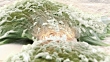
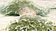
Dividing cancer cells. Coloured scanning electron micrograph (SEM) of colorectal cancer cells undergoing cytokinesis (cell division). Cytokinesis occurs after nuclear division (mitosis), which produces two daughter nuclei. The cells are still attached by a cytoplasmic bridge (centre). Magnification: x3500 when printed at 10 centimetres wide.


ライセンスの購入
商品の使用用途、使用期間、使用国・地域等の条件に基づいたライセンス。
詳細
クレジット:
クリエイティブ写真番号:
618321795
ライセンスタイプ:
ライツレディ
コレクション:
Image Bank Film
最大ファイルサイズ:
1280 x 720 px - 254 MB
クリップの長さ:
00:00:12:00
アップロード日:
リリース情報:
リリースはありません。
マスター:
QuickTime 8-bit Photo-JPEG HD 1280x720 25p