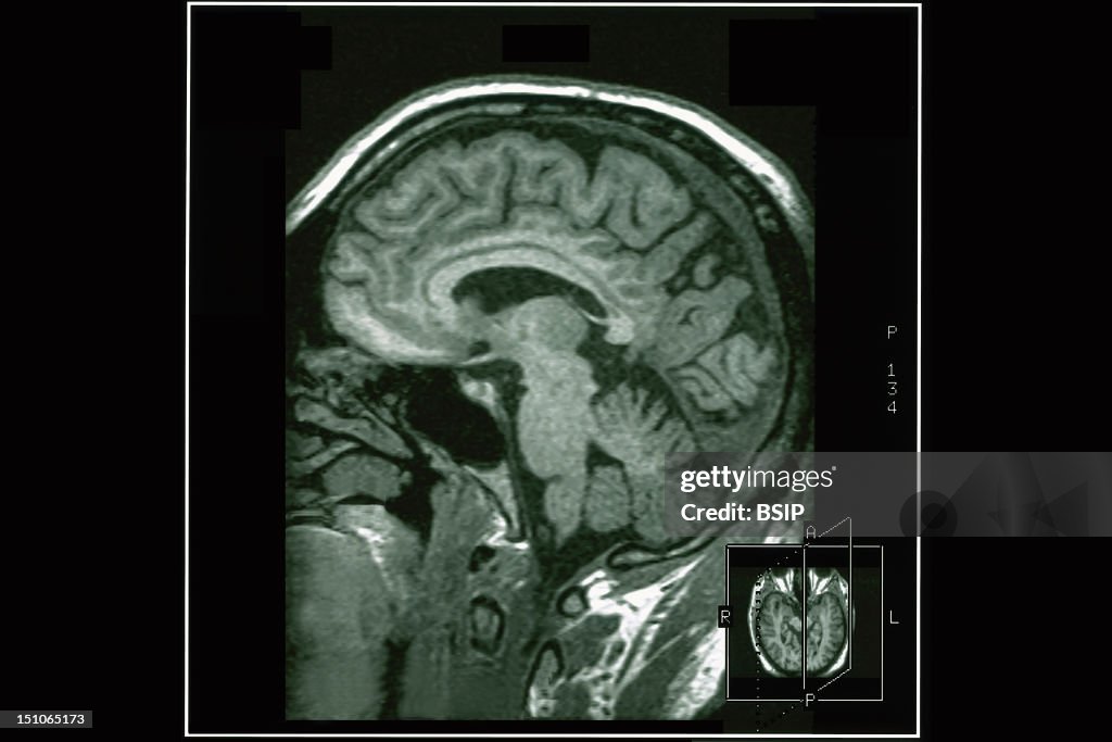
Memory, Mri
Department Of Morphological And Functional Imaging, Led By Professor Fredy, At The Pitie Salpetriere Hospital In Paris, France. Normal Anatomy. Image Of The Last Interconnection In The Memory Process Between The Thalamus And Cingulate Cortex. (Photo By BSIP/UIG Via Getty Images)

ライセンスの購入
どんな用途に素材を使えますか?
¥38,500
JPY
【注意】歴史的な内容の画像は、当時の背景に基づいているため、現在の理解を反映していないテーマや説明が含まれている場合があります。詳細については、こちらをクリックしてください。
詳細
制限:
商業目的またはプロモーション目的で使用する場合は、ゲッティ イメージズのオフィスへお問い合わせください。
クレジット:
報道写真番号:
151065173
コレクション:
Universal Images Group
作成日:
2002年06月13日(木)
アップロード日:
ライセンスタイプ:
リリース情報:
リリースされていません。 詳細情報
ソース:
Universal Images Group Editorial
オブジェクト名:
941_04_0517002
最大ファイルサイズ:
3630 x 2420 px (30.73 x 20.49 cm) - 300 dpi - 2 MB