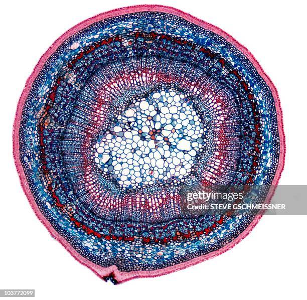
helianthus (sunflower), herbaceous dicot, stem cross section, vascular bundles, cortex, pith. 25x - 顕微鏡図 ストックフォトと画像植物の髄, 維管束, 成長
spinal cord. cross section, 2.5x. shows: gray matter, white matter, dorsal root ganglia, dorsal and ventral roots, central canal, anterior horn cells (motor neuron cell bodies) & meninges - 顕微鏡図 ストックフォトと画像背骨, 正面から見た図
nerve; cross section, myelinated nerve fibers (nerve fibers are dark, surrounded by white myelin sheath), 50x at 35mm. shows a single fasciculus (bundle of nerve fibers), perineurium (blue connective tissue), and endoneurium (blue). - 顕微鏡図 ストックフォトと画像人物
spinal cord cross section, 5x shows: gray matter (golden butterfly), white matter, central canal, dorsal & ventral root, dorsal root ganglion, meninges, dorsal horn, ventral (anterior) horn, & anterior horn cells (motor neuron cell bodies) - 顕微鏡図 ストックフォトと画像顕微鏡, 背骨, ヒトの脳
zea (corn) cross-section. mature stem. scattered vascular bundles. xylem. herbaceous monocot stem, phloem. 5x - 顕微鏡図 ストックフォトと画像顕微鏡, 科学, 拡大イメージ
light micrograph of human sublingual salivary gland; mucous cells and a duct, 100x at 35mm. this is a mixed gland, but is mostly made up of mucous alveoli. - 顕微鏡図 ストックフォトと画像細胞, 唾液, 粘液
This photomicrograph was created from a cultured specimen stained with methylene blue and revealed the presence of a parasitic trophozoite, Entamoeba...
Influenza virus. Viruses of the Orthomyxoviridae family. Avian plague virus. The flu virus is the cause of an infectious and contagious respiratory...
spinal cord cross section. shows gray & white matter, central canal, motor neuron, dorsal and ventral roots. 3x - 顕微鏡図 ストックフォトと画像背骨
Highly magnified, digitally colorized transmission electron microscopic image, showing ultrastructural details exhibited by a single, spherical...ウイルス, コロナウイルス, COVID-19
Highly magnified, digitally colorized transmission electron microscopic image of the particle envelope of a single, spherical shaped, Middle East...コロナウイルス, ウイルス, COVID-19
sperm cells or spermatozoa, immunofluorescent photomicrograph, organs samples, histological examination, histopathology on the microscope - 顕微鏡図 ストックフォトと画像精子, 顕微鏡, 実験室
thymus. 10x shows: numerous lobules, cortex (darker with numerous lymphocytes), medulla (lighter). produces a hormone thymosin. - 顕微鏡図 ストックフォトと画像拡大イメージ, ホルモン, 科学
motor neuron (multipolar) with cell body and many processes (mostly dendrites), spinal cord (magnification x100). this multipolar motor neuron comes from the anterior (ventral) horn of the spinal cord grey matter. the stain is methylene blue and phloxine. - 顕微鏡図 ストックフォトと画像顕微鏡, 細胞, ヒト細胞
microscope of adenoid cystic carcinoma, rare type of cancer exist in many different body sites. - 顕微鏡図 ストックフォトと画像細胞, 抽象的, 科学
leukemia; myeloid, 250x at 35mm. this type of leukemia has its origin in the bone marrow (myeloid tissue). it involves a malignant proliferation of immature white blood cells. this action can crowd out production of rbc s and platelets leading to an - 顕微鏡図 ストックフォトと画像細胞, 白血病, 骨髄組織
cross section of the cerebellum and nerve human under the microscope for education in lab - 顕微鏡図 ストックフォトと画像細胞, 顕微鏡, 筋肉
Influenza virus. Viruses of the Orthomyxoviridae family. Avian plague virus. The flu virus is the cause of an infectious and contagious respiratory...